Ophthalmic Exam
Your horse's eyes should be examined as part of a regular physical exam. However, more thorough testing is needed in the following circumstances:
There is an abnormal appearance to one or both eyes, such as swelling, redness, cloudiness, or discharge
Your horse shows signs of pain, such as holding an eye closed or rubbing at the eyes
You suspect that your horse is experiencing changes in vision (for example, because your horse ran into a fence)
An eye injury has occurred
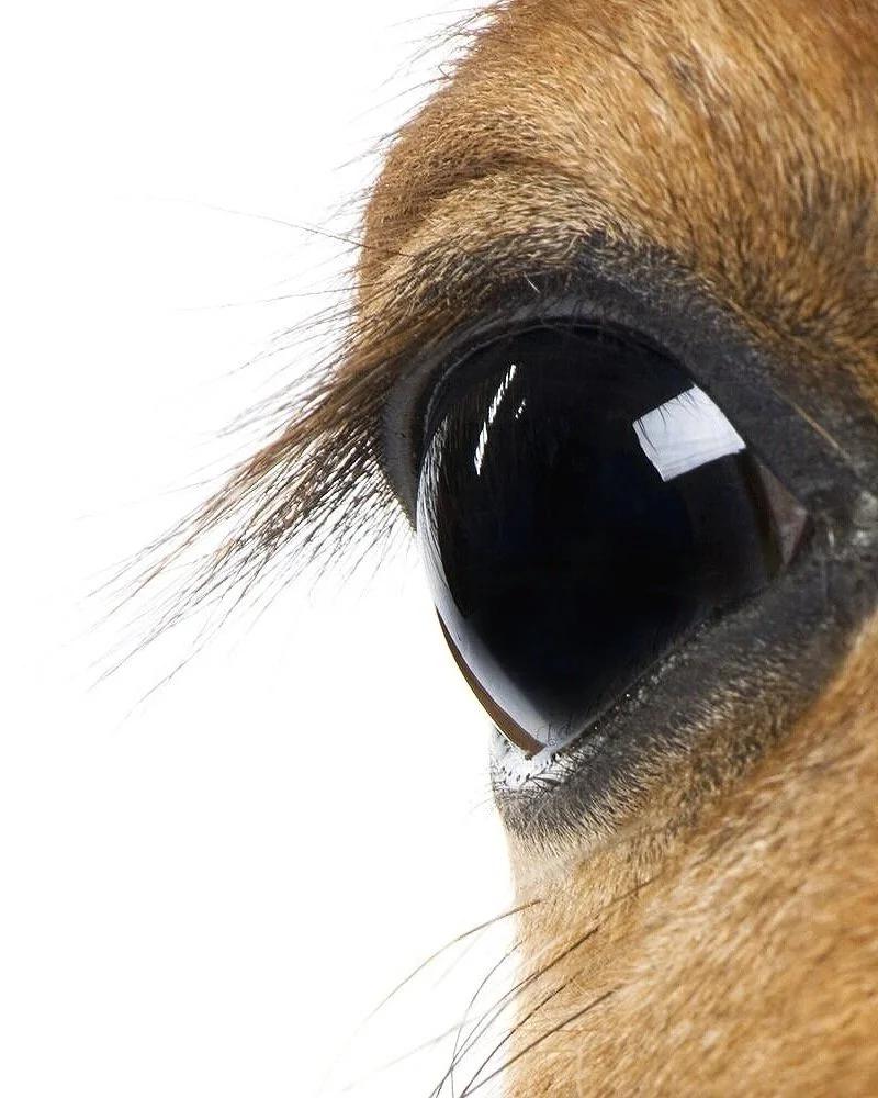
The ophthalmic exam often begins with an evaluation of the horse's vision. Performing the ophthalmic examination in a darkened, quiet area is ideal for evaluating the horse's eyes. A menace test may also be conducted to see if the horse blinks when a finger is moved toward, but without touching, the eye. A pupillary light reflex test is used to evaluate the retina (the sensory membrane that lines the eye's interior), the muscles controlling the iris (the colored portion of the eye), the nerves, and the part of the brain that controls visualization. A bright light is shown into each eye to evaluate both eyes for pupil constriction. An ophthalmic exam usually includes a thorough evaluation of the outer eye structures, including the tissues around the eyes, the eyelids, the duct where the tears drain from the eyes, and the nerves that affect the eyes. At the same time, the eye will be examined for inflammation and infection as well as for foreign bodies and unusual growths.
Because of their large, protruding eyes, horses often inadvertently scratch their cornea (the clear layer on the front of the eye). Because these painful abrasions or ulcers are not always visible with the unaided eye, a fluorescein stain test will be conducted to display the location and size of the abrasion.
Another painful condition for horses is equine recurrent uveitis, which is also known as moon blindness. This condition is due to inflammation of the eye's interior. Certain breeds are predisposed to developing this disease and if not treated it can lead to blindness.
Other diseases, such as glaucoma, can occur in horses. Glaucoma occurs when the fluid pressure inside the eye is too high. This condition is painful and can lead to blindness. If necessary, we can check the pressure of each eye by gently tapping the eye surface with a special instrument that looks like a pen, called a Tonopen. Before testing the eye pressure we place a few ophthalmic anesthetic drops in the eye to numb the eye surface. High pressure is a sign of glaucoma, while low pressure may be a sign of uveitis (inflammation inside the eye).
Corneal Ulcers & Abrasions

Every ulcer in a horse is potentially serious and sight threatening as the equine cornea appears prone to develop infection and heals slowly with extensive scarring. All corneal ulcers, regardless of size or depth, should be considered emergencies, and the patient should receive prompt, aggressive treatment and follow-up care. The risk of bacterial, fungal or mixed infections is quite high in the horse following damage to the corneal epithelium. Corneal ulcerations will generally be painful and the horse will exhibit extreme sensitivity to light, involuntary tight closure of the eyelid, and increased tearing.
Equine Recurrent Uveitis (Moon Blindness)

Equine recurrent uveitis (ERU), also known as moon blindness, has often been cited as the most common cause of blindness in horses. Equine recurrent uveitis is a syndrome that involves all aspects of the equine eye. ERU is characterized by recurrent episodes of intraocular inflammation separated, in most horses, by periods of remission (“quiescence”) in which there are no signs of active intra-ocular inflammation.
Blocked Nasolacrimal Duct (Tear Duct)

The nasolacrimal drainage apparatus of the horse may become obstructed due to a foreign body, calcification, dentition abnormalities, inflammation/infection, or neoplastic processes. In general, tear production in horses is robust, and copious reflex tearing can occur in response to ocular irritation or pain.